Das Röntgen ist ein unverzichtbares diagnostisches Hilfsmittel im Umgang mit einem Wildvogelpatienten und ermöglicht in den meisten Fällen eine schnelle, sichere Diagnose. Eine Sedation des Patienten ist meistens nicht erforderlich (außer bei der Panorama-Aufnahme). Erfahrung des Tierarztes beim Röntgen von Vögeln und Kleintieren ist Voraussetzung für die Anfertigung eines aussagekräftigen Röntgenbildes!
Bei Vorliegen von Frakturen, Luxationen und anderen schweren Verletzungen sollte abgewogen werden, ob dem Patienten die Schmerzen einer betäubungslosen radiologischen Untersuchung zuzumuten sind. In jedem Fall orale Analgetika (Meloxicam) verabreichen! Soll im Anschluss eine operative Wund- oder Frakturversorgung in Allgemeinanästhesie erfolgen, liegt es nahe, auch die Röntgenaufnahme bereits in Narkose anzufertigen. Dabei muss bei allen traumatisierten Patienten eine genaue Abwägung hinsichtlich der Narkosefähigkeit erfolgen. Mauersegler mit Verletzungen, die bereits bei klinischer Untersuchung eindeutig eine Wiederherstellung der Wildbahnfähigkeit ausschließen, werden, sofern aus wissenschaftlichem Interesse eine Röntgenaufnahme vonnöten ist, erst in Narkose oder postmortal radiologisch untersucht.
Wird noch nicht digital, sondern nach alter Methode mit Röntgenkassette gearbeitet, sollte die Kassetten direkt auf den Röntgentisch gelegt werden, um den Abstand vom Objekt zum Film möglichst gering zu halten. Der Abstand Film – Fokus sollte bei 100 cm betragen. Röntgenkassetten der Größe 13 x 18 cm, bei Panorama-Aufnahmen 18 x 24 cm, können benutzt und bei Bedarf durch Ausfokussieren in zwei Hälften unterteilt werden. Abhängig vom verwendeten Röntgengerät haben sich Belichtungwerte zwischen 42 KV/10 mAS und 42 KV/12,6 mAS bzw. zwischen 45 KV/8 mAS und 50 KV/12 mAS als geeignet erweisen. Für die Röntgenaufnahmen von Patienten der Mauerseglerstation wurde in früheren Jahren ein Film-Folien-System aus grünempfindlichem medizinischen Röntgenfilm der Firma Agfa-Gevaert, „Perutz Radiolix G Plus“ verwendet, und um die Strahlendosis zu verringern und kürzere Belichtungszeiten zu erzielen, kamen Verstärkerfolien zum Einsatz. Inzwischen wird digital geröntgt.
Indikation
In folgenden Fällen ist eine radiologische Untersuchung des Mauerseglerfindlings eine notwendige Indikation und muss deshalb durchgeführt werden:
- bei Mauerseglern mit sichtbar asymmetrischer Flügelhaltung und / oder inkongruentem Flügelschlag,
- bei Mauerseglern mit makroskopisch und / oder palpatorisch erkennbaren Defekten der Flügel oder des Schultergürtels (Instabilität, Krepitation, Umfangsvermehrung, Hämatom, offene Wunden),
- bei Mauerseglern mit Lahmheiten, Lähmungen, Versteifungen der Flügel, die sich bei der klinischen Untersuchung nicht klassifizieren lassen.
In folgenden Fällen erscheint eine radiologische Untersuchung des Findlings hilfreich und sollte nach Möglichkeit durchgeführt werden:
- bei Mauerseglern mit schlaffer Lähmung der Beine,
- bei flüggen Jungseglern, die am Boden aufgefunden wurden,
- bei Jungseglern mit dem Vorbericht schwerer und / oder länger andauernder Fehlernährung.
Mauersegler, bei denen der erfahrene Tierarzt bereits beim klinischen Untersuchungsgang eindeutig, offenkundig und ohne die Notwendigkeit weiterführender Untersuchungen eine Wiederherstellung der Wildbahnfähigkeit ausschließen kann, sollten nicht mehr intra vitam geröntgt werden, um ihnen sinnloses Leiden zu ersparen.
Röntgentechnik
Für die radiologische Untersuchung von Mauerseglern haben sich folgende vier Aufnahmearten bewährt:
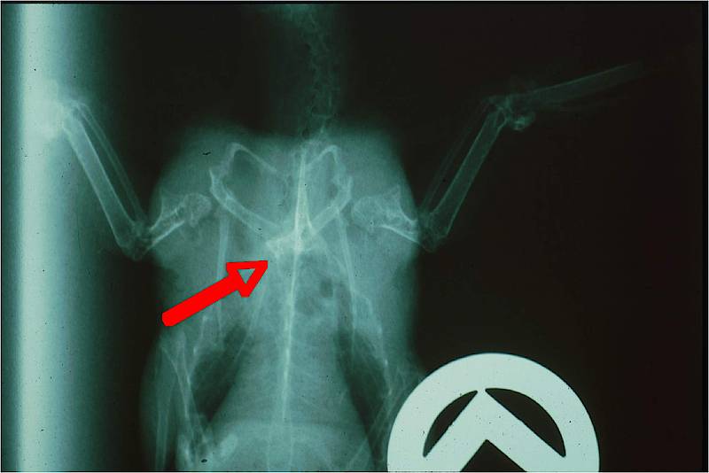
1) Die dorso-ventrale Übersichtsaufnahme
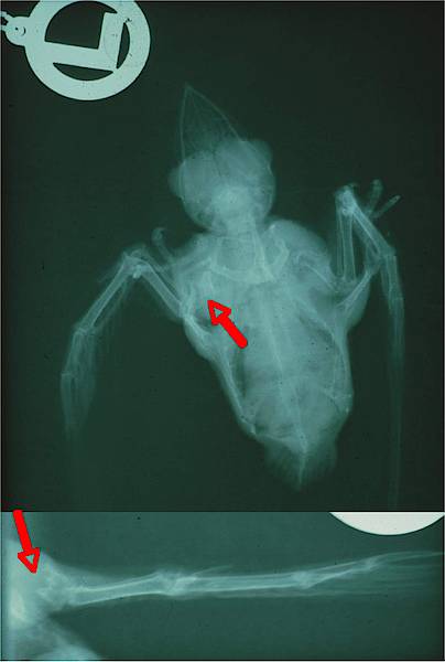
Diese in der Mauerseglerklinik seit 1999 selbst erprobte, speziell für den Mauersegler geeignete und sehr bewährte Projektion wird angewendet, um augenfällige Defekte des Schultergürtels ohne jede Fixation und ohne Stress für den Vogel darzustellen. Der Strahlengang verläuft dorsoventral.
Die Übersichtsaufnahme, in vorliegender Form wahrscheinlich nur beim Mauersegler einsetzbar, ermöglicht eine schnelle und stressfreie Beurteilung des Schultergürtels, ohne dass der Vogel durch Zwangsmaßnahmen beunruhigt wird. Eine Kassette des Formats 13 x 18 cm, mit Seitenzeichen versehen, wird voll ausgeblendet, mit einem Stück Zellstoff oder Küchenpapier bedeckt und der Mauersegler zwanglos daraufgesetzt. Meistens genügt das Überraschungsmoment, wenn der Vogel neugierig umherblickt, um eine dorso-ventrale Übersichtsaufnahme zu erstellen, deren perfekte Lagerung sich aus der natürlichen Haltung des in Brustlage sitzenden Mauerseglers von selbst ergibt. Schultergürtelfrakturen sind auf diese Weise meist sehr gut ersichtlich, Extremitätendefekte aufgrund möglicher Überlagerung mit den Füßen nicht immer zweifelsfrei beurteilbar.
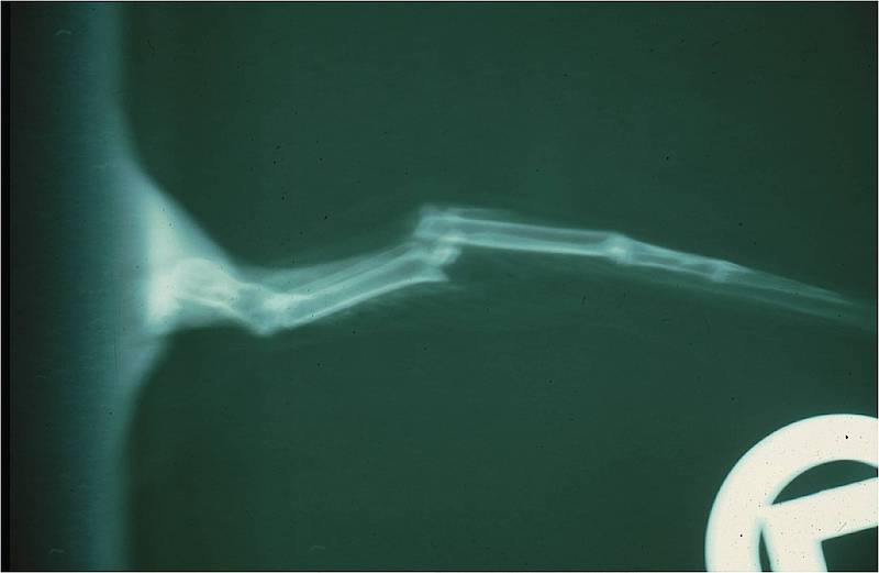
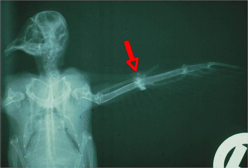
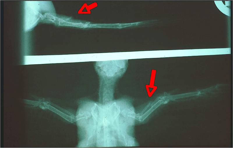
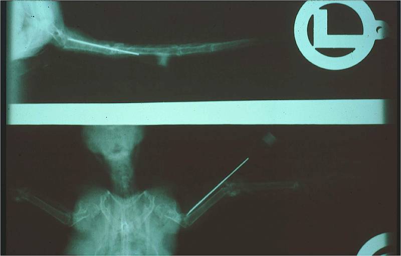
2) Gliedmaßendarstellungen in den beiden Standardebenen
Bei einer radiologischen Untersuchung der Flügel ist die Anfertigung von zwei Aufnahmen, die zueinander in einem Winkel von 90° stehen, obligat und für eine Diagnosestellung absolut unumgänglich. Sofern Röntgenaufnahmen der Beine erforderlich erscheinen, wird ebenso verfahren. Aufnahmen des Flügels werden immer in medio-lateralem und caudo-cranialem Strahlengang erstellt, Aufnahmen der Beine analog in ventro-dorsalem und in medio-lateralem Strahlengang.
Gliedmaßendarstellungen in den beiden Standardebenen können vom Untersuchenden allein am unsedierten Mauersegler vorgenommen werden. Bei der medio-lateralen Aufnahme des Flügels wird der Vogel mit einer Hand über der Brust fixiert, die andere streckt die betroffene Extremität maximal. Anschließend wird auf der zweiten Hälfte der Platte die caudocraniale Ebene projeziert. Dazu hält der Untersuchende den Vogel kopfüber und senkrecht über die Platte, so dass dieser, den Kopf in den Nacken gebogen, mit der Kehle aufliegt. Der betroffene Flügel wird mit der zweiten Hand gestreckt und so dicht wie möglich über der Platte gehalten.
Analog verläuft die Darstellung der Hintergliedmaße, allerdings wird hier auf eine manuelle Streckung der betroffenen Extremität verzichtet, da der Lauf des Mauerseglers zu kurz ist, um die Finger des Untersuchenden außerhalb des Zentralstrahls zu plazieren. Frakturen sind in der Regel auch bei nicht abduzierter Gliedmaße gut zu erkennen. Falls eine Streckung dennoch nötig ist, kann sie mittels eines um den Lauf geschlungenen und gestrafften Gummibandes erfolgen.
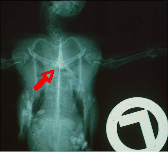
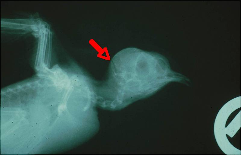
3) Aufnahmen in ventro-dorsal und latero-lateral
Diese radiologische Standarduntersuchung beim Ziervogelpatienten ist beim Mauersegler nur selten erforderlich. Sie dient der Untersuchung und Beurteilung der inneren Organe. Die Kassette wird senkrecht zum Untersucher auf den Tisch gelegt. Auf der unteren Hälfte wird die ventro-dorsale, auf der oberen die latero-laterale Aufnahme angefertigt. Zur Beurteilung von Schultergürteldefekten ist die latero-laterale Aufnahme überflüssig. Notwendig ist eine ventro-dorsale Projektion des Schultergürtelbereiches mit maximal gespreizten Schwingen. Im Bedarfsfall wird die zweite Hälfte der Kassette für eine Speziallagerung zur Abklärung einer Luxation der Articulatio sternocoracoidea benötigt.
Bei der ventro-dorsalen Aufnahme wird der unsedierte Vogel in Rückenlage auf die ausfokussierte untere Hälfte der Kassette gelegt. Mit der linken Hand wird der Kopf fixiert, die rechte Hand streckte die Füße. Anschließend wird das Tier für die latero-laterale Aufnahme auf der nun ausfokussierten oberen Hälfte der Kassette in die linke Seitenlage verbracht. Beide Flügel werden übereinandergelegt und nach dorsal abgestreckt. Mit der Handkante der linken Hand hält der Untersuchende die Flügel auf der Kassette und umfasst zugleich mit Daumen und Zeigefinger den Kopf. Die rechte Hand zieht die übereinanderliegenden Füße in ventro-caudale Richtung.
Für eine ventro-dorsale Aufnahme mit maximal ausgebreiteten Flügeln ist die Hilfe einer zweiten Person nötig. Wieder wird der Vogel in der oben beschriebenen Weise in Rückenlage fixiert. Die zweite Person, die dem Untersuchenden gegenüber auf der anderen Seite des Röntgentisches steht, ergreift nun möglichst weit proximal gleichzeitig beide Flügel und streckt sie maximal.
Eine Speziallagerung kann zur Abklärung einer Luxatio articulatio sternocoracoidea hilfreich sein: Dabei wird nach der ersten Aufnahme mit abduzierten Flügeln eine zweite auf dem oberen Teil der Kassette angefertigt, wobei der Bereich des Schultergürtels eng fokussiert wird. Der Untersuchende fasst den auf dem Rücken gelagerten Vogel an den Karpalgelenken und schiebt beide Flügel nach medial und kaudal zugleich gegen den Körper. Die hierdurch erzielte Hebelwirkung disloziert ein vom Sternum luxiertes Coracoid, in normaler ventro-dorsaler Aufnahme oft nicht sichtbar, deutlich über die Mediane. Beide Aufnahmetechniken sind mit einiger Übung rasch und sicher am unsedierten Segler durchzuführen.
4) Die Panorama-Aufnahme
Sie diente einer Gesamtübersicht der inneren Organe und des Bewegungsapparates in ventro-dorsaler Lagerung bei ausgebreiteten Flügeln und ist nur beim sedierten oder toten Mauersegler möglich. Benötigt wird eine Kassette im Format 18 x 24 cm. Diese Aufnahmeart ist nur sehr selten erforderlich und im täglichen Umgang mit dem Mauerseglerpatienten von geringer Relevanz.