1) Verletzungen des Schultergürtels
Bei den meisten Defekten, die den Schultergürtel betreffen, ist die Prognose infaust und keine Therapie möglich. Klinisch liegt bei Schultergürteldefekten fast immer eine Asymmetrie des Schulterbereiches vor: Eine Schulter "hängt" gegenüber der Gegenseite deutlich tiefer, die Spitze des dazugehörigen Flügels steht höher als die andere. Legt man den Segler probeweise auf den Rücken, vermag er sich aus eigener Kraft nicht wieder herumzudrehen (Drehprobe).
Coracoidfrakturen sind häufige Unfallfolge bei Altseglern, die Flugfähigkeit ist kaum je wiederherzustellen. Durch zumeist mittel bis hochgradige Dislokation der Frakturenden kommt es überdies zu Verletzungen des umliegenden Weichteilgewebes, oft auch der Lunge, wobei perakut Dyspnoe und Blutungen aus dem Schnabel auftreten. Ruhigstellung des Patienten kann zum Verheilen der Fraktur führen, doch sind fast immer Verkürzungen und eine veränderte Statik des Schultergürtels die Folge. Das würde zwar einem Vogel, der nicht auf gutes Flugvermögen angewiesen ist, das Weiterleben ermöglichen, nicht aber einem Mauersegler (seltene Ausnahmen bestätigen die Regel, in vereinzelten Fällen verheilt eine Coracoidfraktur ohne Dislokation, asymmetrische Flügelbewegungen und sichtbar eingeschränktes Flugvermögen werden nicht beobachtet, nach ausgiebiger Physiotherapie kann eine erfolgreiche Freilassung erfolgen).Eine chirurgische Versorgung des Coracoids durch Marknagelung gelingt nicht, da der kurze, kräftige und schwer zugängliche Knochen von umfangreichen Muskelzügen überdeckt wird und ein Zugang ohne erhebliche Verletzung des Weichteilgewebes nicht möglich ist.
Aussichtslos sind auch Gabelbeinfrakturen. Eine Ruhigstellung ist erfahrungsgemäß in allen Fällen erfolglos, eine chirurgische Versorgung nicht möglich, da die Clavicula als filigrane Spannfederkonstruktion nicht stabil zu fixieren ist. Stets kommt es zu Verlagerungen und einer irreversiblen Aufhebung ihrer Funktion.
Scapulafrakturen können in wenigen günstigen Fällen ausgeheilt werden, wenn die Frakturenden nur geringgradig verlagert sind, dem Rippenkorb parallell aufliegen und der betroffene Mauersegler absolute Boxenruhe toleriert. Weist hingegen ein Frakturende nach ventral und lag hochgradige Dislokation vor, kommt es zu umfangreicher Kallusbildung, die betroffenen Mauersegler lahmen fortan und erlangen ihr Flugvermögen nicht zurück. Vermutlich führen starker Druck oder Verwachsungen des Kallusgewebes mit dem Rippenkorb zu erheblicher Bewegungseinschränkung.
Alle Luxationen und Subluxationen im Bereich des Schultergürtels sind aussichtslos und Anlass zur sofortigen Euthanasie. Jeder Bandabriss führt zum Funktionsverlust und nachfolgender Versteifung des betroffenen Gelenkes. Das Flugvermögen wurde beim Mauersegler in keinem Fall wiedererlangt.
Anders verhält es sich mit gedeckten Weichteiltraumata (Prellung). Das klinische Bild mit hochgradiger Lahmheit und Funktionsausfall kann einen Defekt des knöchernen Bewegungsapparates vortäuschen, zumal der verletzte Mauersegler sich auch nicht aus eigener Kraft wieder aus Rückenlage herumdrehen kann (Drehprobe). Ist jedoch radiologisch kein Defekt festzustellen, kann man von einer Prellung ausgehen; die Prognose ist zweifelhaft bis günstig. Zwar kommt es in wenigen Fällen infolge einer schweren Prellung zur Versteifung des involvierten Gelenks und zum dauerhaften Verlust der Flugfähigkeit. Meistens jedoch sind nur 8 – 10 Tage Boxenruhe erforderlich, bis der Vogel den betroffenen Flügel allmählich wieder zu bewegen beginnt. Mit täglicher Physiotherapie, die passives Strecken und Bewegen des Flügels, Massagen, dann Bewegungsübungen in einer Gardine und schließlich Flugübungen in einem geeigneten mit Gardinen abgehängten Trainingsraum umfasst, wird die Flugfähigkeit innerhalb von zwei bis sechs Wochen wiederhergestellt. Ohne ausreichende tägliche Übungen hingegen kommt es zu progressiven Rückschritten und fortschreitender Versteifung des Flügels. Eine manifeste Kontraktur im Schultergelenk kann nicht wieder rückgängig gemacht werden, die Euthanasie ist dann unvermeidlich.

2) Flügelverletzungen
Die Prognose bei Flügelfrakturen richtet sich nach Lokalisation, Alter und Ausmaß der Schädigung. Nicht therapierbar sind Humerusfrakturen. Konservative Frakturversorgung führt zu Verkürzungen und Kontrakturen im Schulter- und / oder Ellbogengelenk. Ein chirurgischer Eingriff ist nicht möglich, da der kurze Humerus körpernah unter umfangreichen Muskelzügen liegt und keinen Zugang gestattet. Sein Condylus dorsalis ist so schmal und dicht am Ellbogengelenk gelegen, dass im Gegensatz zu anderen Vogelarten selbst bei maximaler Beugung kein Austritt eines Marknagels ohne Affektion und spätere Ankylose des Gelenkes möglich wäre.
Prognostisch günstiger sind Frakturen des Unterarms. Beim Mauersegler ist nicht grundsätzlich chirurgischer oder konservativer Versorgung von Radius- oder Ulnafrakturen der Vorzug zu geben. Vor- und Nachteile sind im Einzelfall abzuwägen, vor allem im Bereich der Vordergliedmaßen ist die Anfertigung von Röntgenaufnahmen in beiden Standardebenen unerlässlich! Chirurgische Zugänge und Operationstechniken sowie Spezialverbände beim Vogel (z. B. Figure-eight-Bandage) sind hinlänglich beschrieben (GYLSTORFF und GRIMM, 1987; OROSZ et al., 1992; MARTIN und RITCHIE, 1994; BENNETT, 1997; McCLUGGAGE, 1997) und beim Mauersegler in fast allen Fällen anwendbar. Gewisse Probleme bei der chirurgischen Versorgung von Unterarmfrakturen resultieren aus der Kürze des Oberarms und damit verbunden aus Behinderungen durch den Rumpf beim Reponieren von Frakturen und beim Positionieren des Marknagels. Auch besteht erhöhte Gefahr von Kontrakturen bereits durch geringgradige Gelenksaffektionen. Leicht kann es auch zu Subluxationen während der Reposition der Frakturenden kommen, insbesondere in den Articulationes radiocarpalis und humeroradialis. Die bei anderen größeren Vögeln relativ unkomplizierte Marknagelung von Unterarmfrakturen ist also beim Mauersegler mit gewissen Risiken verbunden.
Radiusfrakturen sind fast immer gedeckt. Klinisch liegt keine Asymmetrie der Flügel vor, jedoch einseitige Lahmheit und meist ein Hämatom auf der Flügelinnenseite. Sind die Frakturenden stark disloziert und stehen nicht mehr in Verbindung, ist eine Marknagelung notwendig, um vollständige Flugfähigkeit wiederherzustellen. Der Nagel, zweckmäßigerweise eine 0,4 mm-Kanüle, tritt bei maximal gebeugtem Karpalgelenk aus dem freien distalen Radiusende an der Articulatio radiocarpalis aus, wird 2 mm über der Haut abgesetzt und 10 Tage belassen. Bei nur geringgradiger Dislokation kann Boxenruhe zu einer befriedigenden Knochenheilung führen. Doch ist zu beachten, dass bei einer frischen Fraktur möglicherweise ein umfangreiches Hämatom die Frakturenden kurzfristig in Position hält, die sich nach dessen Resorption noch erheblich gegeneinander verschieben können. Das Anlegen eines Verbandes ist bei Radiusfrakturen unnötig, da die intakte Ulna als natürliche Schiene fungiert.
Bei Ulnafrakturen ist in der Regel eine chirurgische Versorgung durch Marknagelung (geeignet ist eine 0,5 mm-Kanüle) vonnöten, da es sonst zu erheblichen Verkürzungen und umfangreicher Kallusbildung kommen kann und ein vollständiges Wiedererlangen der Flugfähigkeit nicht gegeben ist. Allerdings besteht aufgrund der durch den austretenden Marknagel bedingten Gelenksaffektion die Gefahr einer Ankylose und späteren Lahmheit.
Bei nur geringgradiger Dislokation der Frakturstücke ist daher abzuwägen, ob eine konservative Versorgung nicht das geringere Risiko darstellt. Dazu ist eine Ruhigstellung unter Verband für ca. 10-12 Tage nötig. Anschließend ist eine mehrwöchige Rekonvaleszenz mit intensiver täglicher Physiotherapie nötig, um befriedigende Flugfähigkeit wiederzuerlangen, da die immobilisierten Gelenke unter Verband sehr schnell versteifen.
Besonders kompliziert und oft nicht befriedigend therapierbar sind Radius-Ulna-Frakturen, die schon klinisch sofort durch Krepitation, starke Hämatombildung, freie Beweglichkeit an der Frakturstelle und asymmetrische Flügelhaltung (Flügel "hängt nach unten") zu diagnostizieren sind. Bei offenen Frakturen 2. und 3. Grades, Splitter- und Trümmerfrakturen sollte aufgrund der schlechten Prognose kein Versuch einer chirurgischen Versorgung unternommen, sondern der Vogel eingeschläfert werden. Ferner muss abgeklärt werden, ob außer der offenkundig sichtbaren Fraktur möglicherweise auch noch eine Luxation vorliegt, die jeden Therapieversuch von vornherein zunichtemachen würde.
Lautet die Prognose zweifelhaft bis günstig, ist eine chirurgische Versorgung unverzichtbar. Eine befriedigende Reposition ist nur durch Marknagelung und das zusätzliche Anlegen eines geeigneten Verbandes (Figure-eight-Bandage) für wenige Tage möglich. Die Marknägel werden 2 mm über der Haut abgesetzt und 12 - 14 Tage belassen. Dabei tritt der Nagel des Radius am Karpalgelenk, der Ulna am Ellbogengelenk aus und kann, wenn die Fraktur stabil ist, ohne weitere Narkose gezogen werden. Zu Komplikationen kann es leider relativ häufig kommen, wenn auch unter Verband keine ausreichende Ruhigstellung der Fraktur gelingt und Scherkräfte auf die Frakturenden einwirken, und wenn die austretenden Marknägel insbesondere im Ellbogengelenk zu Gelenksaffektionen führen, die eine spätere Bewegungseinschränkung zur Folge haben können. Wildbahnfähigkeit ist dann möglicherweise nicht mehr zu erreichen.
Frakturen der Mittelhand haben meistens eine infauste Prognose, da es sich fast immer um offene Splitterfrakturen mit hochgradiger Verletzung, Zerreißung und Kontamination des umgebenden Weichteilgewebes handelt. Bei solchen Frakturen 2. und 3. Grades wird erfahrungsgemäß selbst bei sofortiger Wundversorgung unter Allgemeinanästhesie und Anlegen eines Verbandes die Flugfähigkeit nicht wiedererlangt.
Lediglich bei frischen einfachen Stückfrakturen, gedeckt oder offen 1. Grades, ist eine Therapie fast immer erfolgreich. Es muss sorgsam abgewogen werden zwischen einer Ruhigstellung mittels Figure-eight-Bandage - die stets unter Allgemeinanästhesie erfolgen muss, um die Frakturenden sauber reponieren, mögliche Wunden vernähen und den Verband mit gepolsterter Schiene exakt anlegen zu können! - und einem chirurgischen Eingriff in Form einer intramedullären Nagelung.
In der Mauerseglerstation wird der Versorgung mit Schiene und Verband der Vorzug gegeben, weil sich erwiesen hat, dass der Heilungsverlauf schneller und unkomplizierter vonstatten geht als bei einem operativen Eingriff. Ist dieser dennoch angezeigt, erfolgt der Zugang bei gedeckter Fraktur von medial, bei offener an der Wundstelle. Ein passgerecht zugeschnittenes, steriles Stück Glasfiber wird unter maximaler Beugung beider Frakturenden und Schonung des umliegenden Feingewebes in die Markhöhle des Karpus eingeführt. Über dem Marknagel werden die Frakturenden zusammengeschoben, der Wundbereich gespült, die Hautwunde durch Knopfhefte verschlossen. Für 10 Tage wird eine Figure-eight-Bandage angelegt. Eine knochengängige Antibiose zur Vorbeugung einer Osteomyelitis (z. B. Clindamycin) ist ratsam. Der Nagel verbleibt in der Markhöhle. BENNETT (1997) weist auf die Möglichkeit hin, ein Knochenimplantat zu belassen, falls es nach Verheilen der Fraktur nicht entfernt werden kann. Implantate aus Glasfiber werden in der Literatur nicht erwähnt, sind jedoch in Einzelfällen bereits erprobt worden (N. KUMMERFELD, unveröff. Mitt., 2003).
Beim Vogel werden auch Implantate aus Polyethylen und Polymethyl-methacrylat verwendet, die ebenfalls in der Markhöhle belassen werden können (MARTIN und RITCHIE, 1994). Die Anwendung dieses Materials ist beim Mauersegler bislang nicht möglich, da keine Implantate des benötigten geringen Durchmessers (0,8 - 1,2 mm) verfügbar sind, Glasfiberstäbe hingegen passend zurechtgefeilt werden können. Plattenosteosynthese und externe Fixation sind beim Mauersegler nicht anwendbar, da die knöchernen Strukturen zu klein sind.
Fingerfrakturen sind sehr selten. In der Mauerseglerstation war bei einer Stückfraktur der Phalanx distalis digiti majoris eine konservative Versorgung mit einem schmalen Verband erfolgreich, der den Finger an den Kielen der 7. und 8. Handschwinge fixierte; eine Figure-eight-Bandage war nicht vonnöten.
Nach Ausheilung einer konservativ oder chirurgisch versorgten Flügelfraktur ist unbedingt intensive tägliche Physiotherapie nötig, nicht nur um die volle Funktionsfähigkeit des Flügels zurückzugewinnen, sondern auch um einer Inaktivitätsatrophie der Flugmuskulatur vorzubeugen. Nach Radiusfrakturen ist die volle Flugfähigkeit in der Regel nach circa 3 Wochen wiederhergestellt, bei Radius-Ulna- und Handfrakturen kann es 4 bis 6 Wochen dauern.
Luxationen und Subluxationen der Flügelgelenke sind beim Mauersegler in allen Fällen aussichtslos und Indikation zur Euthanasie. Sie führen unausweichlich zu Kontrakturen und damit zum Verlust der Flugfähigkeit.
Weichteiltraumata müssen von Fall zu Fall nach Ausmaß der Schädigung beurteilt werden. Kleinere, oberflächliche Riss- und Schürfwunden haben bei fachgerechter Wundversorgung eine günstige Prognose, sofern es nicht zur Verletzung darunterliegender Nerven und Sehnenzüge gekommen ist. Selbst unbedeutend erscheinende Platzwunden sollten genäht werden, um ein weiteres Aufreißen der Haut und Austrocknen des Weichteilgewebes zu verhindern. Bei tieferen Wunden ist unter Allgemeinanästhesie sorgfältig zu eruieren, wieweit das darunterliegende Gewebe und auch die Kiele der Hand- und Armschwingen betroffen sind. Oft kann erst nach der Wundtoilette und dem Abheilen der Verletzungen beurteilt werden, ob die Funktionsfähigkeit des Flügels erhalten ist. Skalpierungswunden, bei denen nicht nur Hautanteile, sondern auch Federanlagen zerstört worden sind, haben sich als aussichtslos erwiesen. Mit mehr als einer fehlenden Handschwinge pro Flügel kann ein Mauersegler nicht mehr als wildbahnfähig eingestuft werden.
Weichteiltraumata des Flügels sind nicht selten mit schwerer Beschädigung einer oder mehrerer Handschwingen verbunden. Die Federn können geknickt oder abgebrochen sein. Vom Ziehen einer defekten Handschwinge ist dringend abzuraten, da es danach sehr häufig zu erheblichen Störungen beim Wachstum einer neuen Feder kommt oder gar keine Feder mehr nachgeschoben wird. Traumatisch oder mechanisch bedingte Großgefiederschäden sollten immer durch die falknerische Methode des Schiftens behoben werden, die sich in allen bisherigen Fällen gut bewährt hat.
Bislang beobachtete degenerative oder vermutlich metabolisch bedingte Veränderungen im Flügelskelett hatten meist eine zweifelhafte Prognose, müssen aber nach dem Bild beurteilt werden, das der betroffene Vogel über mehrere Wochen bei der Physiotherapie und im Flugtraining bietet. Liegen dystrophische Veränderungen von Gelenkflächen vor, ist die Prognose infaust. Bei Verbiegungen langer Röhrenknochen, mehrfach bei Radius und Carpometacarpus aufgetreten, zeigen die meisten betroffenen Jungsegler so gutes Flugvermögen, dass es gerechtfertigt erscheint, sie unter Kontrolle freizulassen.
Bei klinischen Bildern, die auf eine Lähmung hindeuten, sollte abgewartet werden und der Vogel über mehrere Wochen täglich mehrmals Physiotherapie erhalten. Ein Jungvogel mit einseitiger Flügellähmung nach intramuskulärer Injektion in den Brustmuskel war erst nach 8 Wochen intensiven Trainings wieder flugfähig.
3) Verletzungen des Rumpfes
Frakturen im Bereich des Rumpfes sind selten. Da sie schwerwiegend sein können und unter Umständen schwierig zu diagnostizieren sind, sollte jedoch gerade bei ungeklärter Flugunfähigkeit, Dyspnoe und Tachykardie das Rumpfskelett besonders aufmerksam radiologisch untersucht werden. Vollständige Sternumfrakturen fallen möglicherweise bei der klinischen Untersuchung zunächst nicht auf, betroffene Segler zeigen allerdings die o. a. Symptome. Erst die radiologische Untersuchung in ventro-dorsaler und latero-lateraler Projektion zeigt eine Verschiebung in der Linie der Carina sterni, wenn Brustbeinkiel und Sternum quer gebrochen sind. Daraus resultiert nicht nur Flugunfähigkeit insbesondere durch die Instabilität des Rumpfskeletts, sondern vor allem bei jeder Bewegung des Vogels Druck des frakturierten Sternums auf das unmittelbar anliegende Herz und Verlagerung von Herz und Leber nach dorsal. Wegen hochgradiger Allgemeinstörungen müssen solche Patienten euthanasiert werden.
Bei einer unvollständigen Sternumfraktur mit hochgradig disloziertem Riss in der Carina sterni gelang in der Mauerseglerstation die chirurgische Reposition. Der Mauersegler erlangte nach mehrwöchiger Physiotherapie sein Flugvermögen vollständig zurück und konnte freigelassen werden. Eine mehrfache Rippenfraktur wurde einmal infolge eines Katzenbisses beobachtet. Trotz umgehender chirurgischer Wundversorgung konnte der Altsegler nicht gerettet werden.
Für Weichteiltraumata gelten dieselben Bedingungen wie im Abschnitt "Flügelverletzungen" aufgeführt. Unbedingt sollte jeder noch so kleine Riss lege artis versorgt und genäht werden, da die Haut über der massiven Pectoralismuskulatur sehr straff aufliegt und leicht weiter einreißen kann, während im Rückenbereich bereits harmlos anmutende Verletzungen die Rippen und die darunter liegenden Lungen betreffen können, weil die Haut sehr dünn ist und kein Schutz durch weiteres Weichteilgewebe besteht.
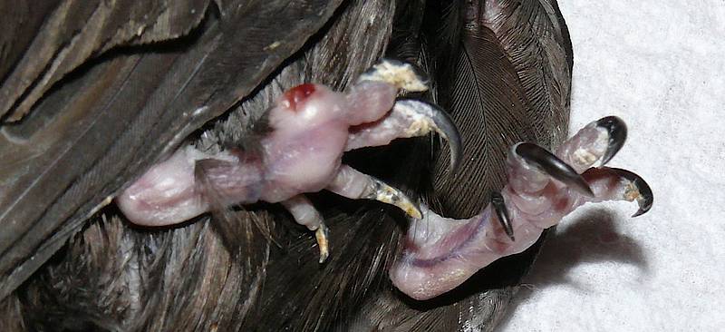
4) Verletzungen der Hintergliedmaßen
Bei Frakturen und Luxationen der Hintergliedmaßen ist die Prognose ist meist günstig. Im Gegensatz zu vielen anderen Vogelarten stellt Funktionseinschränkung oder gar Verlust einer Hintergliedmaße das Überleben eines Mauerseglers in freier Natur vermutlich nicht in Frage. Jedenfalls kommen sogar einbeinige Mauersegler jahrelang in freier Natur zurecht (E. KAISER, unveröff. Mitt., 2001).
Einfache und doppelte Beinfrakturen – meistens ist der Tibiotarsus betroffen - treten sehr häufig beim Absturz sehr junger Segler aus dem Nest auf, wenn der Nestling noch weitgehend unbefiedert und zugleich wohlgenährt, also schwer ist, und den Absturz noch nicht durch sein Großgefieder abmildern kann. Altvögel hingegen verfangen sich oftmals am Nistplatz oder bei der Nistplatzsuche mit den Füßen in Fäden und hängen hilflos vom Dach. Sofern sie gerettet werden, sind strangulierte, luxierte und frakturierte Hinterextremitäten die Folge. Ein Röntgenbild ist für Diagnosestellung und Therapie in der Regel nicht nötig, meistens ist die klinische Untersuchung ausreichend für das weitere Vorgehen.
Bei komplizierten Splitterfrakturen, mehrfacher Luxationen und Nekrosen nach Strangulation sollte das betroffene Bein amputiert werden. Bewährt hat sich das Absetzen im Knie- oder im Intertarsalgelenk. Zu berücksichtigen ist die wesentlich stärkere Beinmuskulatur eines Altseglers im Gegensatz zum Jungsegler, es kann zu profusen Blutungen kommen, wenn Muskeln und Gefäße nicht sorgsam ligiert werden.
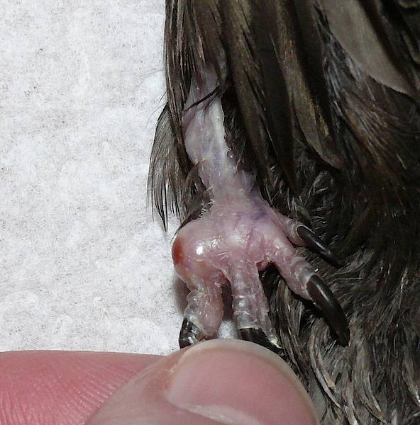
Bei unkomplizierten Frakturen des Tibiotarsus ist in den meisten Fällen eine kurzfristige Fixation mit einem Körperverband ("Leibbinde") erfolgreich. Frakturen bei Nestlingen verheilen meist schon nach wenigen Tagen. Etwaige Dislokation und geringgradige Fehlstellung des Fußes sind zu tolerieren.
Pododermatitiden, meist Sekundärbefunde, gelegentlich selbst verursacht durch Verletzungen mit den eigenen Krallen, haben sich beim Mauersegler als kompliziert und weitgehend therapieresistent erwiesen. Medikamentelle Behandlung wie auch chirurgische Eröffnung der Ballenabszesse bleiben weitestgehend erfolglos. Nur in wenigen Fällen bei fehlernährten Jungseglern lagen eitrige Abszesse vor, bei betroffenen Altseglern waren es eher diffuse Schwellungen des Weichteilgewebes. Da ein verlängerter stationärer Aufenthalt wegen der erhöhten Belastung, der die Füße des Mauerseglers durch die liegende Körperhaltung ausgesetzt sind, kontraindiziert erschien, wurden die betroffenen Altvögel nach Ausheilung ihrer primären Defekte freigelassen.

5) Verletzungen des Kopfes und Halses
Relativ häufig haben Mauersegler Unterschnabelfrakturen. Seltener betroffen sind Altvögel, bei denen die Verletzung gelegentlich als Unfallfolge nach einem Anflug auftritt. Primär ist abzuklären, ob nicht weitere, schwerwiegendere Verletzungen mit infauster Prognose vorliegen, ehe man an die Versorgung einer Schnabelfraktur denkt.Bei Jungseglern - Fundvögeln in Menschenhand - entsteht eine Schnabelfraktur hauptsächlich durch Unachtsamkeit beim gewaltsamen Füttern des Pfleglings. Beim Öffnen des Schnabels wird die filigrane rostrale Spitze der Mandibula abgeknickt, oft beidseitig. Sofern es sich um eine frische, unkomplizierte Fraktur handelt und die Hornscheide des Schnabels unversehrt ist, heilt sie meist ohne weitere Versorgung binnen weniger Tage, wenn das Öffnen des Schnabels zum Füttern fortan mit äußerster Sorgfalt und proximal der Fraktur erfolgt.Liegt eine offene Fraktur mit Dislokation der Frakturenden vor, sind Wundtoilette und das Anlegen einer Schiene ratsam. Die Versorgung sollte gegebenenfalls unter Allgemeinanästhesie vorgenommen werden, um die Schiene akkurat plazieren zu können. Bewährt hat sich ein längs aufgeschnittenes Stückchen Federkiel, z.B. von einer Taube, das über die Frakturstelle geschoben wird. Zu einem Abgleiten der Schiene kann es kommen, wenn sich unter ihr Kallus bildet und eine Aufwölbung entsteht. In der Regel ist dann aber bereits ausreichende Stabilität erreicht.Bei komplizierten, unfallbedingten Frakturen war im Einzelfall auch eine chirurgische Stabilisierung mit Cerclagen aus resorbierbarem Nahtmaterial und anschließender Ruhigstellung erfolgreich. Die Fütterung erfolgte von der anderen Schnabelseite aus unter minimaler Öffnung der Mundspalte. Dabei wurde nicht, wie üblich, der Unterschnabel nach unten, sondern der Oberschnabel nach oben gezogen.
Frakturen oder Luxationen des Zungenbeins können ebenfalls durch Unachtsamkeit beim Füttern durch Laien verursacht werden. Zu solchen Verletzungen des Apparatus hyobranchialis kann es kommen, wenn beim Einführen der Futterinsekten die Zunge versehentlich gewaltsam nach hinten in den Schlund gebogen wird. Schwere rezidivierende Schluck- und Atembeschwerden, schlimmstenfalls Ersticken sind die Folge. Manchmal heilt der Defekt so ab, dass der betroffene Vogel beschwerdefrei leben kann. Bleibt jedoch eine dauerhafte Dyspnoe und / oder Dislokation der Zunge zurück, muss er eingeschläfert werden.
Eine radiologisch dokumentierte Schädelfraktur verlief tödlich: Der adulte Mauersegler mit einer Fissur des Os occipitale und einer schweren Augenverletzung gelangte zwar noch lebend in die Mauerseglerstation, wurde aber wegen hochgradiger Allgemeinstörungen umgehend eingeschläfert. Er blutete aus dem Ohr, pathognonomisch für einen Schädelbasisbruch.
Augenverletzungen haben beim Mauersegler eine schlechte Prognose. Meist sind schwere Anflüge die Ursache, es kommt zu irreversiblen und höchstgradig schmerzhaften Schädigungen wie Linsenabriss, Blutungen in die vordere Augenkammer, Anstieg des Augeninnendrucks und Hornhautdefekten. Während beim Ziervogel die Extirpation eines zerstörten Auges möglich ist, ist ein Mauersegler mit nur einem Auge nicht wildbahnfähig. Halbierung des Sehfeldes, Verlust des sektoral räumlichen Sehens würden Zielorientierung und daher auch den Nahrungserwerb so beeinträchtigen, dass ein Überleben ausgeschlossen ist. Mauersegler mit irreversiblen Augenverletzungen müssen euthanasiert werden. Kann dies nicht unverzüglich vorgenommen werden, ist dringend für ausreichende Analgesie (lokal und systemisch) Sorge zu tragen!
Augenaffektionen können auch durch Fremdkörper verursacht werden, z. B. verklebte Federn, die ins Auge stechen und Reizungen hervorrufen, schlimmstenfalls Hornhautläsionen. Eine erfolgreiche Behandlung ist in vielen Fällen möglich, doch sollte ein Augenspezialist konsultiert werden. Mit antibiotischen Augentropfen ist es nicht (immer) getan, manchmal ist ein Glätten der Hornhaut unter Lokalanästhesie nötig. Komplikationen sind nicht selten. Niemals unbedacht Kortikoide lokal applizieren, bei perforierenden Hornhautverletzungen können sie die Entstehung eines Ulcus provozieren!
Weichteiltraumata im Kopfbereich umfassten Skalpierungswunden, z. B. durch Streifen von Drähten im Flug, sowie sekundär Verletzungen durch einen Beutegreifer, z. B. durch eine Elster, wenn der Segler nach einem Anflug niedergegangen war. Je nach Ausdehnung und Tiefe der Wunde muss umgehend eine Wundversorgung erfolgen, gegebenenfalls unter Allgemeinanästhesie. Liegen Teile der Schädeldecke frei, ist dafür zu sorgen, dass die Wunde feucht gehalten wird, bis der Defekt geschlossen und eine Wundnaht angelegt werden kann. Verletzungen im Kehl- und Halsbereich können Pfählungs- oder Risswunden sein, wie im Fall eines Altseglers, der mit gefülltem Kehlsack einen Nagel streifte. Die Kehle war von der rostralen Unterschnabelspitze bis zum Halsansatz perforiert. Umgehende chirurgische Wundversorgung und das Anlegen einer doppelten Naht waren erfolgreich.